
Illuminate
what matters
Clarity. Speed. Confidence.
High-performance multiplex immunofluorescence reagents designed for robust, scalable, and practical tissue workflows – spatial biology at the ease of standard IHC.
What matters
to you?
Revolune delivers a reagent system designed to remove friction - so you can focus on discovery, not on decoding protocols.
Revolune simplifies multiplex IF without the compromises.
From assay transferability to reproducibility and clinical relevance, Revolune removes the barriers that have traditionally limited multiplex IF adoption.

Assay Transferability
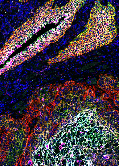
Transfer existing TSA assays with confidence: Direct comparisons to customer TSA workflows show preserved and in some cases improved marker detection.
Why it matters: It combines ease of adoption and workflow compatibility and signal performance, all while preserving validated biology.

Strong concordance with established IHC workflows
Revolune multiplex IF demonstrates strong concordance with traditional DAB staining, enabling a seamless transition from conventional IHC into multiplex imaging.
Why it matters: Transfer of existing antibody concentrations from DAB to mIF. Faster assay development with minimal re-optimization. Greater confidence in biological interpretation. Easier adoption of multiplex workflows using already validated assays.

Speed & Throughput
Cut workflow time nearly in half - because staining and amplification happen in parallel, total assay runtime can be reduced by roughly 50% compared to traditional cyclic TSA workflows.
Why it matters: Faster turnaround. Higher throughput. Better scalability. Routine multiplex IF should not require multiple days per experiment.

Designed for clinically meaningful insights
Revolune enables researchers to move beyond technical multiplexing and toward biologically actionable understanding. By combining multiplex IF with AI-powered analysis in a streamlined end-to-end workflow, researchers can identify clinically relevant biomarker patterns that support Patient stratification, Translational research, Target evaluation strategies, Deeper understanding of disease biology.
This is more than imaging performance - it’s a pathway from spatial biology to clinical impact.
Illuminate what matters
without the workflow complexity.
We Removed the Pain Points Others Built Around.
Revolune provides a gentler, automation-friendly spatial biology workflow that removes the complexity of traditional multiplex imaging — no stripping, no conjugation, no sequencing optimization, and no specialized imaging lock-ins.
NO STRIPPING.
Preserve tissue integrity with parallel staining and amplification — without harsh cyclic workflows.
NO CONJUGATION.
Use the antibodies you already trust.*
No custom conjugation required.*Initial offering supports rabbit IgG primary antibodies.
NO RIGID PANEL DESIGN.
Mix and match detection signals freely between biomarkers without rebuilding workflows.
NO SEQUENCING OPTIMIZATION.
Forget troubleshooting antibody order and panel logic. Fast assay development becomes simple and repeatable.
Illuminate what matters Out-of-the-box
Revolune reagents are built to simplify spatial biology with parallel staining and amplification, enabling fast, scalable multiplex IF from the start. Designed for reproducibility, flexibility, and real-world workflows, our chemistry enables fast, scalable multiplex IF without the complexity of traditional approaches.
“The reproducibility and multiplexing performance of Revolune reagents supported high-quality spatial biomarker profiling for precision oncology applications.”

Multiplex IF Kits for Every Stage of Discovery
From focused biomarker studies to higher-plex spatial profiling, the Illuminate Reagent portfolio scales with your research needs. Choose from 4-plex to 7-plex configurations and generate high-quality multiplex IF data using the antibodies, workflows, and imaging systems you already trust.