
Frommultiplex images to quantitative, spatial insightwith PhenoScout AI.
Revolune helps researchers generate high-quality multiplex immunofluorescence data without the complexity of traditional mIF workflows. With PhenoScout AI by Mindpeak™, available through Revolune, you get there without becoming an image-analysis expert.
From one image to your whole study
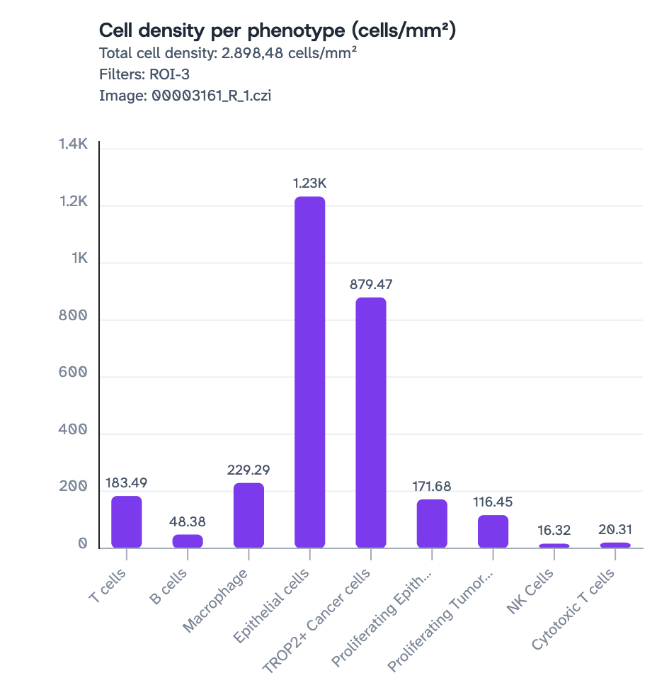
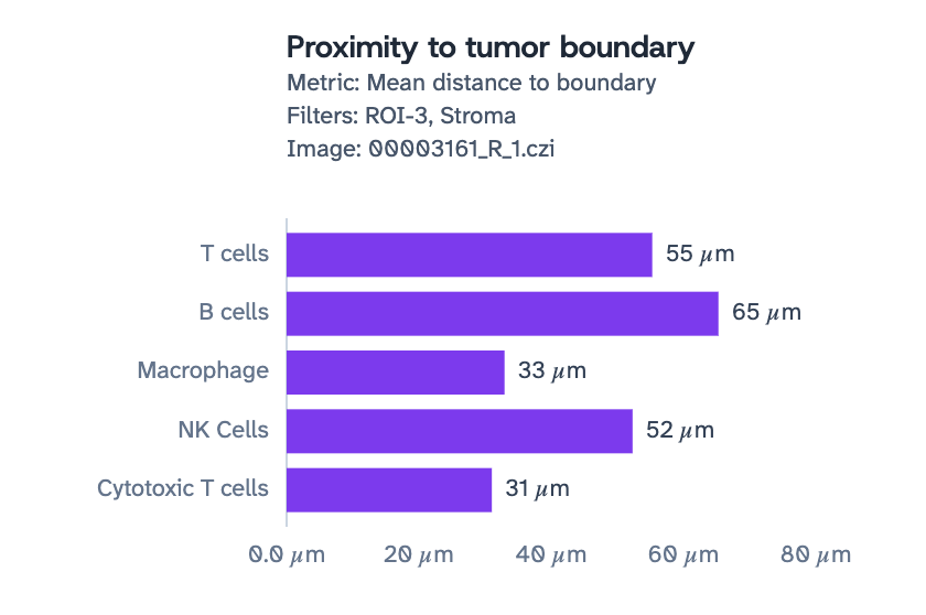
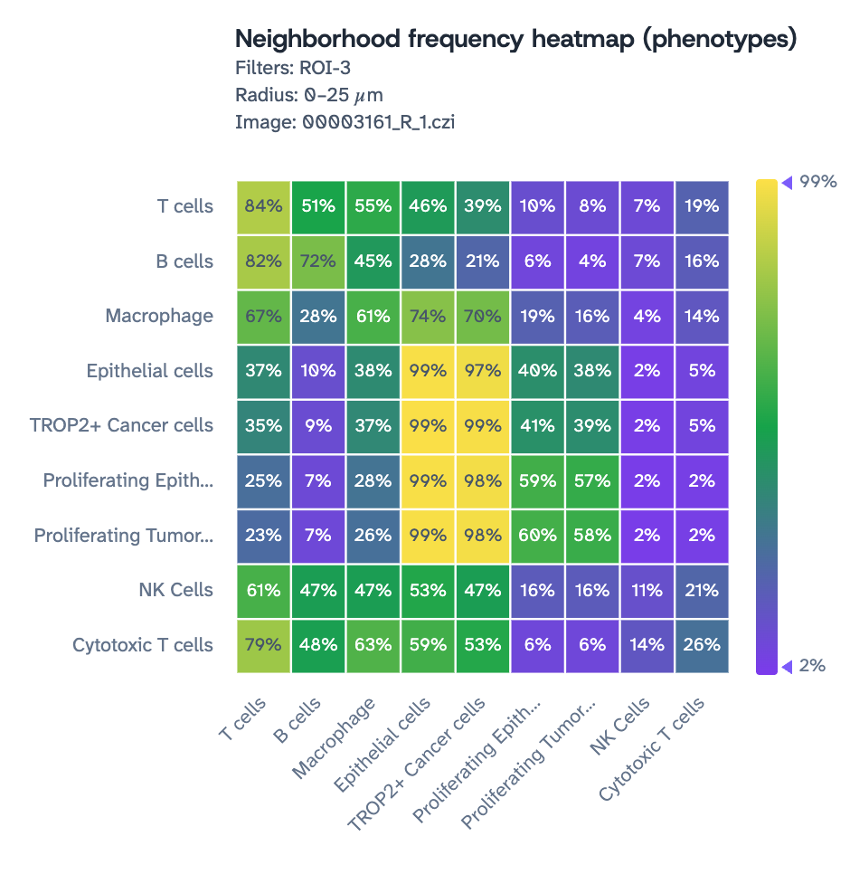
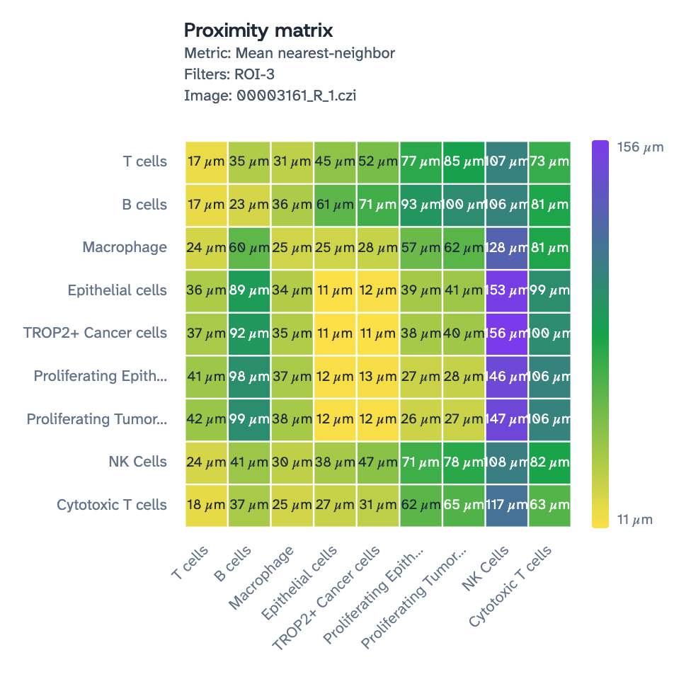
PhenoScout AI phenotypes every cell and keeps going: who is there, who sits next to whom, how far each population stays from the tumor — and any readout you want to build on top. The examples below are a single ROI.
Less complexity. More biology.
Your mIF workflow shouldn't become complicated at the analysis step.
Multiplex immunofluorescence generates rich information about cells, biomarkers and their spatial relationships — but extracting it can demand specialized image-analysis expertise, algorithm development and time-consuming optimization. Pre-trained models automate the key steps, so you move from images to reproducible quantitative data without building a pipeline from scratch.
Simplicity
No image-analysis expertise required
Upload your images and use pre-trained AI models rather than developing and tuning algorithms yourself.
Quantification
Quantify biomarker expression
Generate quantitative marker information at the cellular and subcellular level.
Phenotyping
Automated cell phenotyping
Identify and classify cells across multiplex tissue images to better characterize complex tissue environments.
Spatial biology
Explore spatial biology
Turn multiplex images into data that reveals cell populations, biomarker patterns and spatial relationships within tissue.
With PhenoScout AI analyze what matters to you.
Simpler staining. Simpler analysis. More time for biology.
From staining to insight
Revolune owns the workflow end to end. PhenoScout AI by Mindpeak sits at the analysis step — the same philosophy of simplicity, extended past the image.
01 — STAIN
Multiplex with Revolune
Use the primary antibodies researchers already know and trust.
02 — IMAGE
Capture mIF data
Generate multiplex tissue images using the compatible imaging workflow.
03 — ANALYZE
Apply PhenoScout AI
Use pre-trained models for segmentation, phenotyping and biomarker quantification.
04 — DISCOVER
Focus on biology
Explore spatial readouts, cell populations and marker patterns.
Powerful AI. Without the image-analysis learning curve.
PhenoScout AI is designed to make advanced mIF analysis accessible to research teams without requiring specialized bioinformatics or image-analysis expertise.
Pre-trained AI models
Start analyzing without training your own models.
Cell & tissue segmentation
Identify tissue regions, cells and subcellular compartments.
Phenotype identification and quantification
Characterize cell populations across complex tissue environments.
GxP ready audit trail
Guarantee full data integrity from raw image to final report.
Spatial readouts
Explore how cells, phenotypes and biomarkers are organized within tissue.
Built for mIF research
Analyze multiplex immunofluorescence images using AI developed specifically for tissue analysis.
Inside PhenoScout AI
Click through the readouts your images turn into.

PhenoScout AI
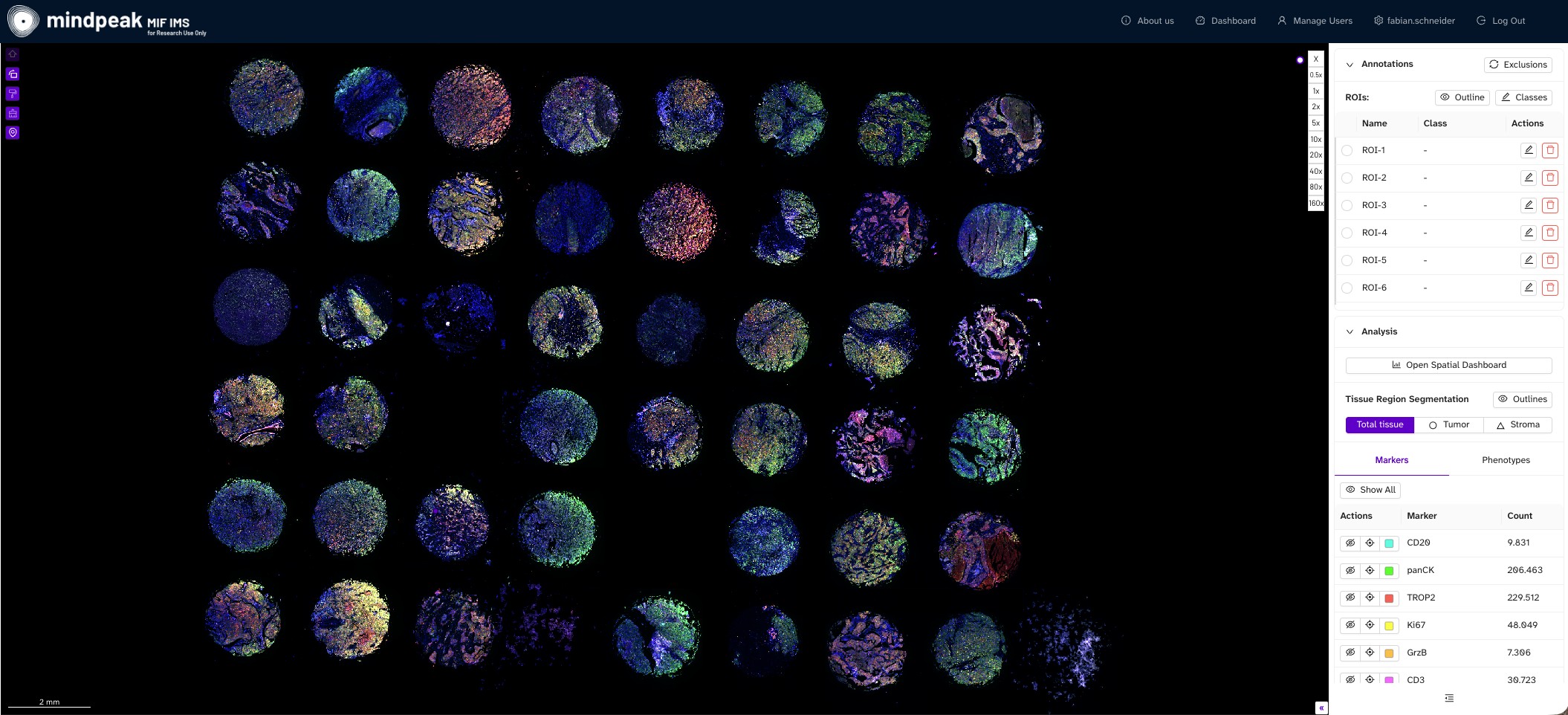
Whole-slide view of a 7-plex breast cancer TMA — every core, one pass, user ROIs listed alongside. (Automated TMA workflow in development)
PhenoScout AI
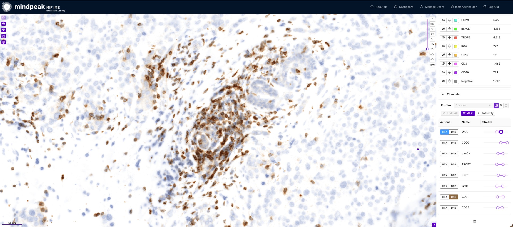
Virtual IHC of CD3 — the familiar DAB brown-and-blue readout, rendered from any mIF channels.
PhenoScout AI
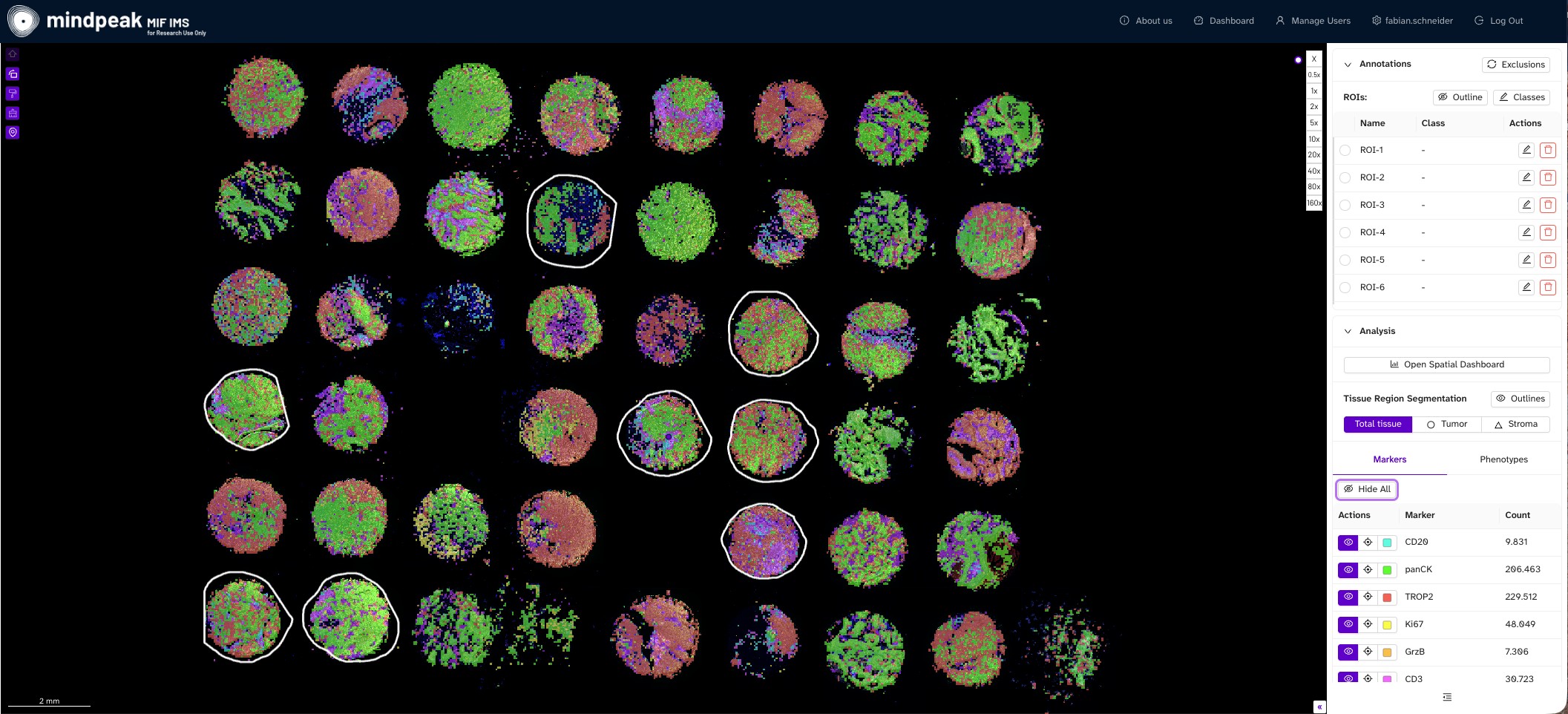
Low-mag marker expression across the tissue image showing the predominant marker per tile in a heat-map overlay
PhenoScout AI
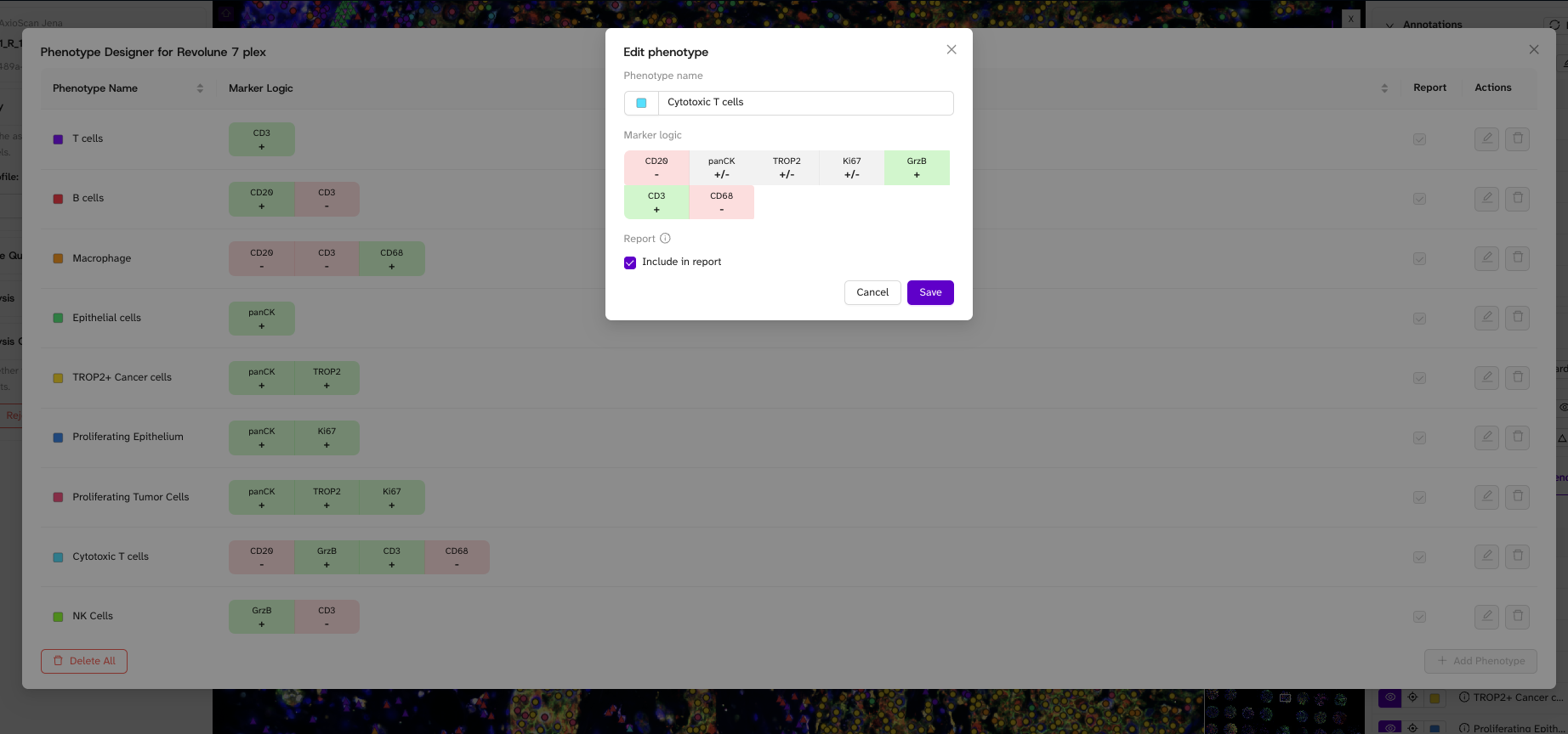
PhenoScout offers a supervised Phenotype designer per assay to setup the outputs you need in an intuitive and fast workflow.
PhenoScout AI
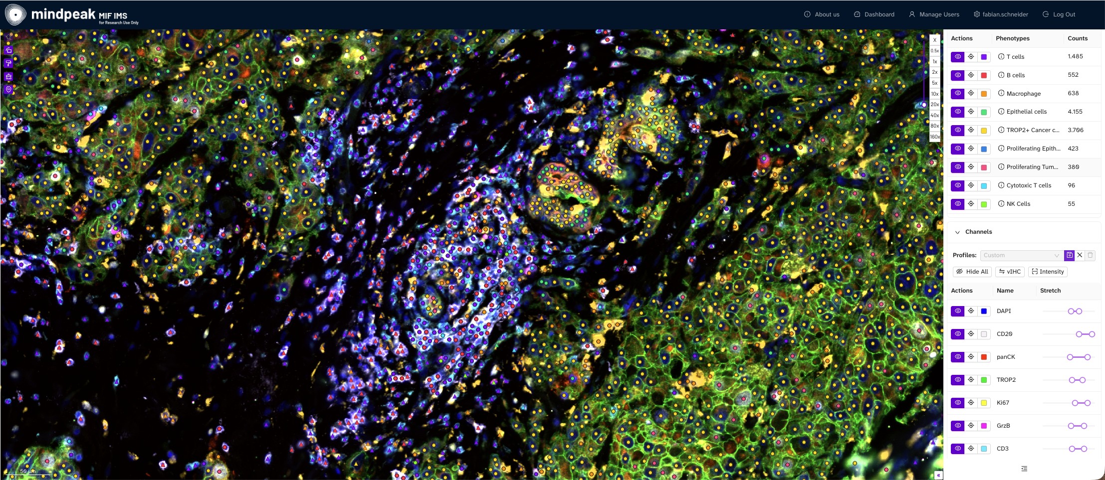
Review Phenotype overlays on cellular level
PhenoScout AI
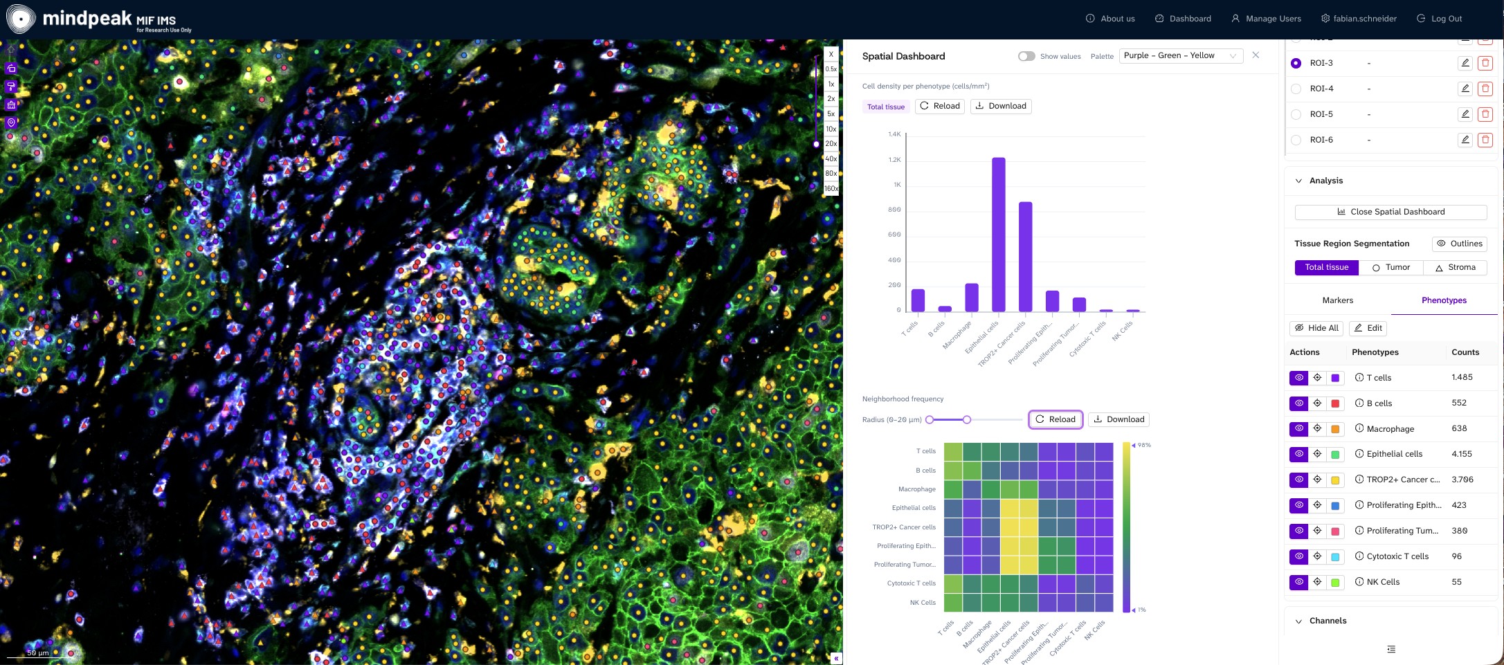
Automated Spatial Dashboard for data insights, from Cell counts and density per Marker and phenotype to neighborhood frequency with user defined radius, proximity analysis and distance to tumor border, all ready to export for publication.
PhenoScout AI
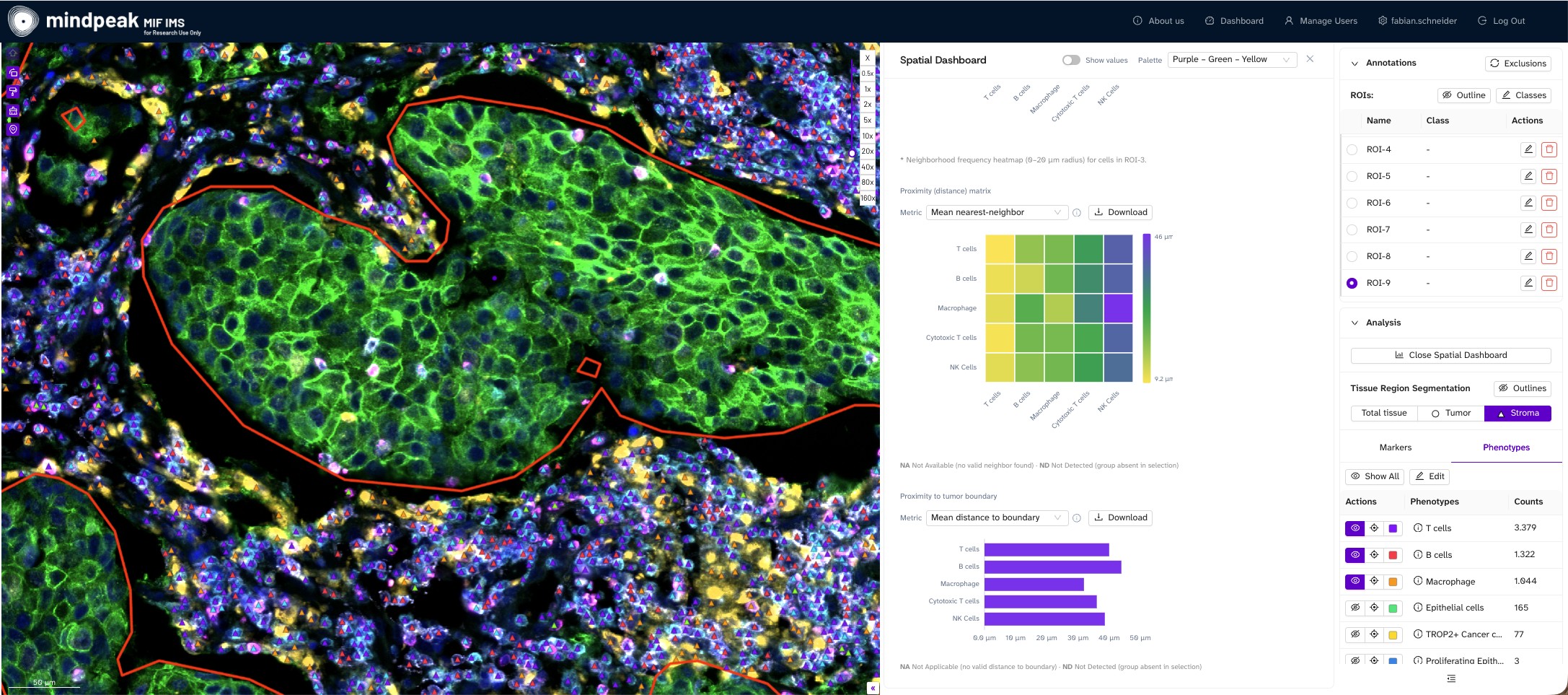
Tumor and stroma outlined automatically — proximity measured against that boundary.
Revolune launch offer
A free PhenoScout AI trial with every Revolune reagent kit.
Buy any Revolune mIF reagent kit and we set you up with a full PhenoScout AI trial — phenotyping, quantification and spatial readouts on your own images, at no extra cost.


See it first
Request a demo
Not ready to order? Walk through PhenoScout AI live with Fabian, or send one of your own multiplex images and get the phenotyped, quantified readout back.
Request a demo

PhenoScout AI is developed by Mindpeak™ and available through Revolune. For Research Use Only. Not for use in diagnostic procedures.
FAQs
Choosing an mIF analysis workflow raises practical questions — inputs, models, deployment and how it fits the lab you already run. Here are the answers we are asked most.
Multiplex immunofluorescence (mIF) and brightfield images, including cyclic stainings of 20+ channels, from all major spatial platforms — RareCyte Orion, Akoya PhenoImager HT, Lunaphore COMET and others. AI analysis currently runs on the PhenoScout mIF model, with a generic brightfield (IHC) model available as an add-on. Support for further modalities is already planned.
No. PhenoScout is a turn-key, off-the-shelf solution. Pre-trained models run on upload, so there is no pipeline to build, no algorithms to tune and no bioinformatics queue to wait in.
Two pre-trained mIF models — mIF-membrane for membrane and cytoplasm markers such as HER2, CD8, panCK and CD68, and mIF-nucleus for nuclear markers such as Ki67 and FOXP3 — plus ready-to-use brightfield IHC-membrane and IHC-nucleus models. All are built on the peakFrontier foundation model, and the mIF model is trained on 30+ immune and cancer biomarkers.
PhenoScout is cloud-native and browser-based, accessible anywhere, anytime. An on-premise deployment (dockerized, in-network) is available on request for teams that need image and result data to stay inside their own infrastructure.
PhenoScout integrates with leading imaging hardware, including the ZEISS Axioscan 7 / SlideStream workflow, and with laboratory IT systems (LIMS / IMS) — from launching results in a separate viewer to embedding them directly in third-party IMS viewers.
PhenoScout AI is For Research Use Only — not for use in diagnostic procedures. It is built to GxP-ready standards, with a full audit trail for every image ensuring traceable, reproducible analysis. Revolune is ISO 13485 (QMS) certified, GDPR-compliant and made in Germany, and develops a separate line of CE-IVD clinical products.