
The Right Plex for Every Research Questions
The Illuminate Reagent Portfolio offers flexible multiplex immunofluorescence solutions from 4-plex to 7-plex. Whether you're validating biomarkers or exploring complex tissue biology, every kit delivers the same streamlined workflow, trusted compatibility, and high-quality results.
Not Sure Which Kit Fits Your Research?
Our team can help you select the optimal plex level based on your biomarkers, assay requirements, and research goals. Let's find the right solution for your workflow.
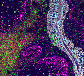
Built for Real-World Spatial Biology
Every Illuminate kit is designed to reduce complexity while delivering the flexibility and performance modern multiplex IF workflows demand.

Use the antibodies you already trust and validate.
.jpeg)
Preserve tissue integrity throughout the entire workflow.

Build multiplex panels faster with fewer compromises.
Stay flexible across instruments, assays, and applications.
FAQs
Choosing the right multiplex immunofluorescence workflow often raises important questions around compatibility, assay development, imaging requirements, and scalability. Explore the answers to the most common questions about Illuminate Reagents and discover how our technology fits into existing spatial biology workflows.
Currently for research use only. However, the workflow is designed to be compatible with clinical lab requirements such as automation and reproducibility.
TSA is effective but involves complex, time-consuming workflows and can damage tissue through repeated cycles. This approach provides comparable sensitivity with a simpler, faster, and less harsh workflow.
A 4-plex kit is available now, with 5- to 7-plex kits coming soon. The DNA-based system is inherently scalable.
Yes, ProLong Gold mounting medium
You need wavelengths of 488,555,647 and 750
A typical 4-plex assay can be completed in approximately 6 hours, significantly faster than traditional multiplex approaches.
No.The system is compatible with standard fluorescence microscopes and existing lab infrastructure. No proprietary hardware is needed.
Highly reproducible.Parallel staining reduces variability, and the workflow is compatible with automation. Fewer steps result in fewer potential sources of error. Furthermore, the stainng protocol is automatable and can be run on an autostainer to increase reproducibility.
Primary antibodies are pre-incubated with Revolune Connectors which act as anchors for signal amplification. A DNA scaffold is then built at each antibody site, allowing multiple fluorescent probes to bind. This results in strong signal amplification while enabling all antibodies to be applied simultaneously in one step.
TSA relies on sequential staining and enzymatic amplification, which increases time and complexity. Cyclic IF requires repeated stripping, which can damage tissue. Revolune enables parallel staining with DNA-based amplification, eliminating stripping and reducing workflow time significantly.
Antibodies are first pre-incubated with Revolune Connectors. After that, all antibodies are applied simultaneously, followed by amplification and detection. This removes the need for iterative staining cycles.
No. The system works with unmodified, pre-validated primary antibodies. Revolune Connector binding is achieved through a simple pre-incubation step, preserving existing validation and workflows.
Yes, within species compatibility.Current kits support rabbit antibodies, with expansion to mouse antibodies soon. No custom conjugation or proprietary antibody panels are required.
No.Existing antibody panels can be reused. There are no strict species pairing constraints, allowing flexible panel design and faster assay development.
DNA scaffold-based amplification.
Each antibody site builds a DNA scaffold that can bind multiple fluorescent probes. This increases signal intensity and improves signal-to-noise ratio.
Signal is generated through DNA-based amplification rather than diffusion-based deposition. This results in low background and high specificity, leading to cleaner staining and improved spatial accuracy.
Amplification is localized at the antibody binding site. The DNA scaffold remains spatially constrained, preserving subcellular resolution.
No.
There is no antibody stripping or harsh treatment involved. This preserves tissue morphology and antigen integrity, which is especially important for sensitive samples.
Still have questions?
Get in touch with our team to discuss your application, research goals, or technical requirements. We're happy to help you find the right solution for your workflow.