
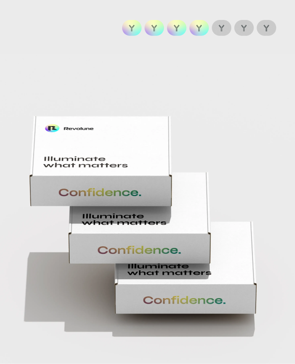
Revolune Illuminate-P4-R
A modular reagent system for flexible multiplex IF assay development. This kit is designed to perform single or multiplex immunofluorescence stainings of FFPE (Formalin-Fixed Paraffin-Embedded) samples.
Details
The user can select up to four different rabbit IgG primary antibodies (Ab). These Ab are pre-labeled with 4 different Connectors (488, 555, 647 & 750). The staining protocol can be performed manually or on the Leica Bond system. The reagents are sufficient to stain 10 slides.
Need More Information?
If you have specific application requirements, need additional information, or simply want to discuss your project before placing an order, we're happy to help. Send us an email or use our contact form and a member of our team will get back to you shortly.
How Illuminate Reagents work
A simple three-step approach designed to make multiplex IF faster, gentler, and easier to adopt. It protects your tissue, accelerates your assay development, reduces your costs, and gives you the clarity to answer the scientific questions that matter most.

Add your antibodies in one step
Use your existing, pre-validated primary antibodies without modification. Simply pre-incubate each antibody with one of our Connectors. Mix your pre-incubated primary antibody-Connector complexes and apply them to the tissue section in a single step.
- Use your trusted antibodies – no conjugation or revalidation
- Adjust panels easily
- Reduce staining time and complexity

Amplify your signal
Our amplification reagents build a DNA scaffold at each of our Connectors.
- Detect low abundance targets
- Increase signal without adding background noise
- Get more data from every tissue section

Detect what matters to you
Fluorescent probes bind to multiple sites on each DNA scaffold. Therefore, many fluorophores are recruited to each antibody site to yield bright staining.
- Produce bright signals for confident analysis
- Work seamlessly with standard fluorescent imaging systems
- Deliver consistent, reproducible results across experiments

Want the technical details?
Download our manual and automation-ready protocols to see how easily Illuminate Reagents can fit into your multiplex IF workflow.
FAQs
Choosing the right multiplex immunofluorescence workflow often raises important questions around compatibility, assay development, imaging requirements, and scalability. Explore the answers to the most common questions about Illuminate Reagents and discover how our technology fits into existing spatial biology workflows.
Currently for research use only. However, the workflow is designed to be compatible with clinical lab requirements such as automation and reproducibility.
TSA is effective but involves complex, time-consuming workflows and can damage tissue through repeated cycles. This approach provides comparable sensitivity with a simpler, faster, and less harsh workflow.
A 4-plex kit is available now, with 5- to 7-plex kits coming soon. The DNA-based system is inherently scalable.
Yes, ProLong Gold mounting medium
You need wavelengths of 488,555,647 and 750
A typical 4-plex assay can be completed in approximately 6 hours, significantly faster than traditional multiplex approaches.
No.The system is compatible with standard fluorescence microscopes and existing lab infrastructure. No proprietary hardware is needed.
Highly reproducible.Parallel staining reduces variability, and the workflow is compatible with automation. Fewer steps result in fewer potential sources of error. Furthermore, the stainng protocol is automatable and can be run on an autostainer to increase reproducibility.
Primary antibodies are pre-incubated with Revolune Connectors which act as anchors for signal amplification. A DNA scaffold is then built at each antibody site, allowing multiple fluorescent probes to bind. This results in strong signal amplification while enabling all antibodies to be applied simultaneously in one step.
TSA relies on sequential staining and enzymatic amplification, which increases time and complexity. Cyclic IF requires repeated stripping, which can damage tissue. Revolune enables parallel staining with DNA-based amplification, eliminating stripping and reducing workflow time significantly.
Antibodies are first pre-incubated with Revolune Connectors. After that, all antibodies are applied simultaneously, followed by amplification and detection. This removes the need for iterative staining cycles.
No. The system works with unmodified, pre-validated primary antibodies. Revolune Connector binding is achieved through a simple pre-incubation step, preserving existing validation and workflows.
Yes, within species compatibility.Current kits support rabbit antibodies, with expansion to mouse antibodies soon. No custom conjugation or proprietary antibody panels are required.
No.Existing antibody panels can be reused. There are no strict species pairing constraints, allowing flexible panel design and faster assay development.
DNA scaffold-based amplification.
Each antibody site builds a DNA scaffold that can bind multiple fluorescent probes. This increases signal intensity and improves signal-to-noise ratio.
Signal is generated through DNA-based amplification rather than diffusion-based deposition. This results in low background and high specificity, leading to cleaner staining and improved spatial accuracy.
Amplification is localized at the antibody binding site. The DNA scaffold remains spatially constrained, preserving subcellular resolution.
No.
There is no antibody stripping or harsh treatment involved. This preserves tissue morphology and antigen integrity, which is especially important for sensitive samples.
Still have questions?
Get in touch with our team to discuss your application, research goals, or technical requirements. We're happy to help you find the right solution for your workflow.