A Flexible Path to Multiplex Immunofluorescence: Discover an Alternative to Cyclic TSA and InSituPlex®
.webp)

Key takeaways:
- The multiplex IF chemistry a lab chooses determines how flexible, reproducible, and vendor-independent the program stays over time.
- Cyclic TSA achieves multiplexing through sequential staining, signal amplification, and stripping rounds, a process that adds tissue stress, runtime, and reproducibility burden with each cycle.
- DNA-barcoded antibody platforms, such as InSituPlex®, remove the secondary antibody step but tie panels to proprietary barcoded primary antibodies and a single vendor's software and analysis ecosystem.
- A Connector-based workflow lets researchers label their own validated primary antibodies and run multiplex IF on a standard fluorescence microscope, adding spatial context without new instrumentation or vendor lock-in.
Introduction
There is more than one way to build a multiplex immunofluorescence (multiplex IF) assay, and the chemistry a lab chooses shapes more than image quality. It shapes flexibility, reproducibility, and how easily a program moves from one study to the next.
This post compares three common multiplex IF methods: cyclic tyramide signal amplification (TSA), DNA-barcoded antibodies (InSituPlex®), and a novel Connector-based workflow that functions as both a TSA alternative and an InSituPlex alternative. For background on what multiplex IF is and why it matters, see our earlier post on moving from IHC to multiplex IF.
What Are Three Ways Labs Build a Multiplex IF Assay Today?
Most multiplex IF programs today take one of three paths:
- Cyclic tyramide signal amplification (TSA staining) multiplexes through sequential rounds of staining, signal amplification, and antibody stripping.
- DNA-barcoded antibody platforms, such as InSituPlex® (ISP), use primary antibodies pre-conjugated to DNA barcodes paired with iterative imaging rounds to decode each marker.
- A Connector-based workflow that labels a researcher's own validated primary antibodies with a fluorescent tag ahead of a single simultaneous staining step.
Each path generates multiplex IF data, but the trade-offs in antibody freedom, workflow time, and vendor dependence differ substantially.
How Does Cyclic TSA Multiplexing Work in Practice?
Cyclic TSA multiplexes by repeating stain, amplify, and strip cycles for each additional marker. Because each cycle strips the previous round's antibody before the next begins, tissues are vulnerable to damage. Each round compounds stress on tissues and adds hands-on time, which can complicate reproducibility across sites. Cyclic TSA processing is subject to higher background noise which can lower sensitivity, especially for low-frequency markers. Longer runtimes and limited scalability are common trade-offs as plex increases.
Where Does InSituPlex® Fit, and What Are the Trade-Offs?
InSituPlex® takes a different approach. Primary antibodies, pre-labeled with DNA barcodes, bind protein targets directly, eliminating the need for secondary detection antibodies. Signal amplification combined with sequential imaging rounds decodes multiple biomarkers from a single staining step, producing quantitative, pathology-grade output.
For translational, biomarker, and CRO teams, the trade-off is antibody access. Panels are typically built from the vendor's barcoded antibody menu, or an existing antibody must be sent out for custom conjugation, which adds time and cost before a study can begin. Panel design, imaging software, and downstream analysis are all proprietary to the vendor's ecosystem. As spatial biology consolidates, illustrated by Ultivue's InSituPlex® platform moving under Vizgen following the two companies' 2024 merger, teams increasingly weigh vendor flexibility and continuity before committing multi-year programs to a single proprietary stack.
How Does a Connector-Based Workflow Compare?
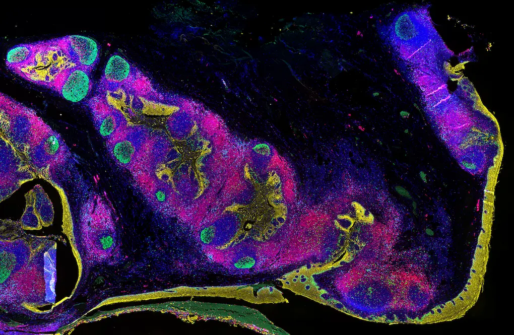
A Connector-based workflow removes several of these constraints. Revolune Connectors label a researcher's own validated IgG primary antibodies directly, so there is no conjugation, no antibody stripping, and no rigid barcoded panel to design around. Antibody sequencing optimization (deciding which primary goes on which round) is unnecessary because all antibodies are applied together in a single, simultaneous staining step. The result is multiplex immunofluorescence without antibody stripping, built on the primary antibodies researchers already trust.
The workflow utilizes a standard fluorescence microscope, so no new imaging hardware is required, and it is compatible with existing automation for labs operating at scale. In practice, this means roughly half the runtime of a cyclic TSA protocol, with strong concordance between multiplex IF results and the DAB IHC results already validated in the lab. Explore Revolune's reagents to see how the chemistry supports this workflow.
Which Multiplex IF Approach Fits Your Program?
The right choice depends on what a program needs from the assay. Highest-plex discovery and tissue atlasing work, where dozens of targets are screened at once, generally favors DNA-barcoded or other high-plex platform approaches built for that scale. Programs that need targeted, reproducible protein panels using antibodies the lab already trusts, with minimal vendor lock-in and no new instrument to purchase, are better served by a Connector-based workflow. The table below compares DNA-barcoded antibody multiplexing (ISP), cyclic TSA, and Revolune’s Connector-based workflow across the criteria that matter most when a program is weighing spatial biology without instrument lock-in against a higher-plex, vendor-defined platform.
This fit matters most when moving from biomarker discovery into validation, where reproducibility and antibody continuity carry more weight than plex count. Programs built around immune cell phenotyping in the tumor microenvironment often prioritize that continuity over plex count. For a closer look at how these criteria play out in practice, see our post on the transition from biomarker discovery to validation.
Ready for a Flexible Multiplex IF Workflow?
Spatial biology at the ease of standard IHC is the goal behind Revolune’s multiplex IF platform. If your team is comparing multiplex IF chemistries for an upcoming study, explore Revolune's technology and reagent portfolio and contact us to discuss your specific project requirements.