From IHC to Multiplex IF: Add Spatial Context Without Rebuilding Your Workflow

Key takeaways:
- Researchers can easily transition from standard IHC to multiplex IF using the validated primary antibodies they already own.
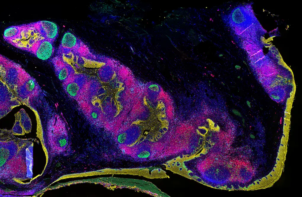
- Multiplex IF preserves intact FFPE tissue architecture while detecting four or more biomarkers simultaneously, delivering the single-cell spatial context that single-plex IHC cannot provide.
- The Revolune workflow eliminates the cyclic staining, stripping, and sequential optimization burden associated with traditional TSA-based multiplex approaches.
- As spatial proteomics and AI-powered tissue analysis continue to reshape oncology and immunology research, multiplex IF enables labs to generate high-quality spatial data without single-source instrumentation or reagent systems.
Introduction
Spatial biology is reshaping how oncology and immunology researchers understand disease. More labs are asking what their tissue sections can tell them about cellular proximity, immune infiltration, and biomarker co-expression. Multiplex immunofluorescence (multiplex IF) is at the center of that shift. But for many researchers already running validated immunohistochemistry (IHC) workflows, the assumption has been that getting there requires new instrumentation, new reagents, and months of re-optimization. That assumption now deserves to be reconsidered.
This post is for researchers considering the move from standard IHC or multiplex IHC to multiplex IF. Whether you are working with FFPE tissue in a translational lab, a biopharma biomarker team, or a core facility, the goal here is to give you an accurate picture of what multiplex IF is, why it matters now, and how the workflow has become more accessible than ever.
What Is Multiplex Immunofluorescence, and How Does It Differ from Standard IHC?
Standard IHC uses a single primary antibody and a chromogenic reporter such as DAB to detect one protein target per tissue section. Multiplex immunohistochemistry (multiplex IHC or mIHC) extends this concept by detecting multiple targets, though often through sequential staining cycles. Multiplex immunofluorescence (multiplex IF or mIF) goes further: it allows simultaneous detection of four or more biomarkers on a single FFPE section using fluorescent reporters, preserving intact tissue architecture throughout.
The key distinction is spatial resolution at the cellular level. With multiplex IF, you can see which cells express which proteins, how those cells are positioned relative to one another, and what that spatial organization means biologically. Single-plex IHC cannot deliver that cellular context. Spatial proteomics, the broader field of measuring protein expression in tissue with spatial resolution, depends on techniques like multiplex IF to generate that data at scale.
Why Does Spatial Context Matter for Oncology and Immunology Research?
Understanding how immune cells and tumor cells interact requires more than knowing which proteins are present. It requires knowing where those proteins are expressed and in what cellular context. The tumor microenvironment is defined by the spatial relationships between immune populations, stromal cells, and tumor cells. Immune cell phenotyping by multiplex IF can reveal whether cytotoxic T cells are actually infiltrating a tumor or being excluded from it, whether macrophage subtypes are polarized toward pro- or anti-tumor states, and how immune checkpoint marker expression varies across tissue regions.
This kind of spatial phenotyping has become central to translational oncology and biomarker development. Regulatory and scientific bodies including the Society for Immunotherapy of Cancer (SITC) have published consensus guidelines underscoring the importance of multiplex tissue imaging for immuno-oncology research, recognizing that single-marker data alone leaves critical biological questions unanswered.1 Demand for reproducible, scalable spatial workflows across multi-site studies is growing, and multiplex IF is the method that answers that demand.
Why Has Multiplex Immunofluorescence Felt Out of Reach Until Now?
The barriers to multiplex IF adoption trace back to a few core technical challenges. First, the same-species problem: standard IHC antibody panels often use primary antibodies from the same host species, making it difficult to distinguish secondary detection without signal crosstalk. Second, many established multiplex workflows rely on cyclic staining approaches, including tyramide signal amplification (TSA staining), which require sequential rounds of staining, signal amplification, and antibody stripping. These workflows are technically demanding, time-consuming, damaging to tissues, and difficult to reproduce across sites.
Add to that the perception that multiplex IF requires specialized imaging hardware, and the result has been a technology that many researchers have followed from a distance rather than adopted in practice. A TSA alternative that reduces workflow complexity without sacrificing data quality is something the field has needed. That is the subject we will address more directly in the next post in this series.
Do You Have to Abandon Your Existing Antibodies and Optimization Work?
No. This is the most important point for labs already running validated IHC panels. The antibodies you have optimized for DAB-based IHC can transfer directly into a multiplex IF workflow. Antibody concentrations validated for chromogenic detection serve as a reliable starting point for multiplex IF titration, reducing the optimization burden significantly. You are extending your existing expertise, not replacing it.
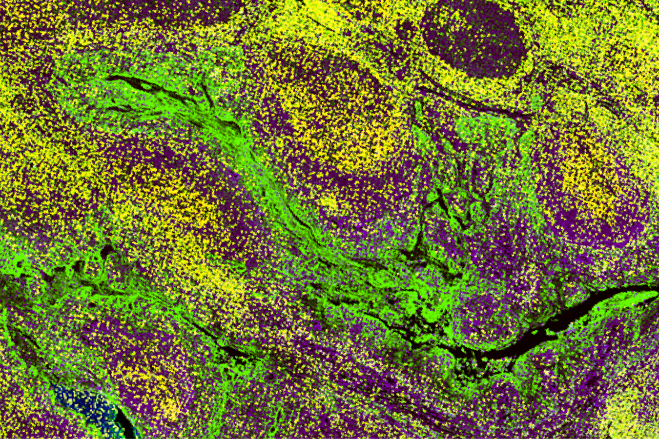
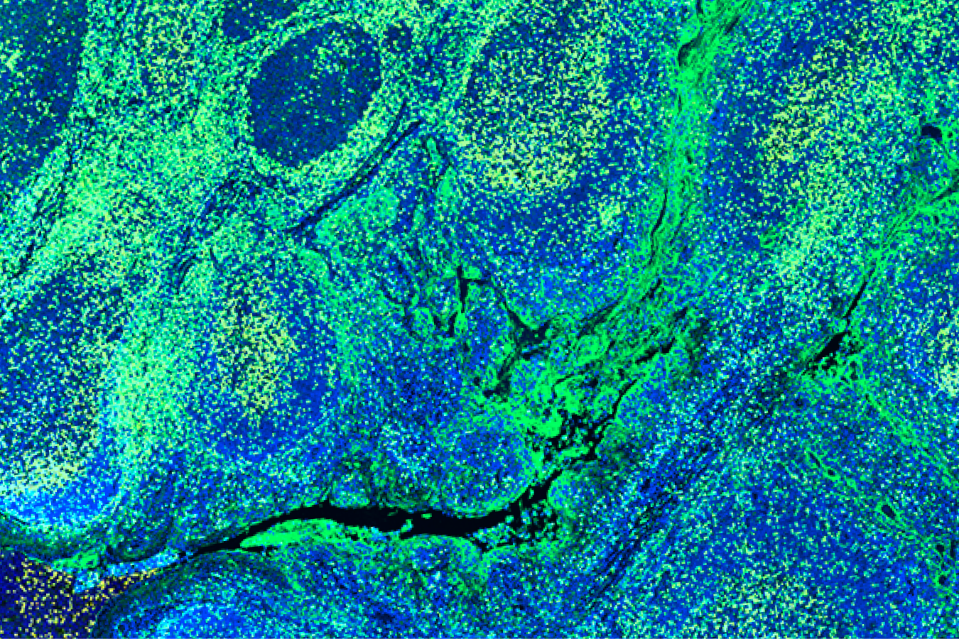
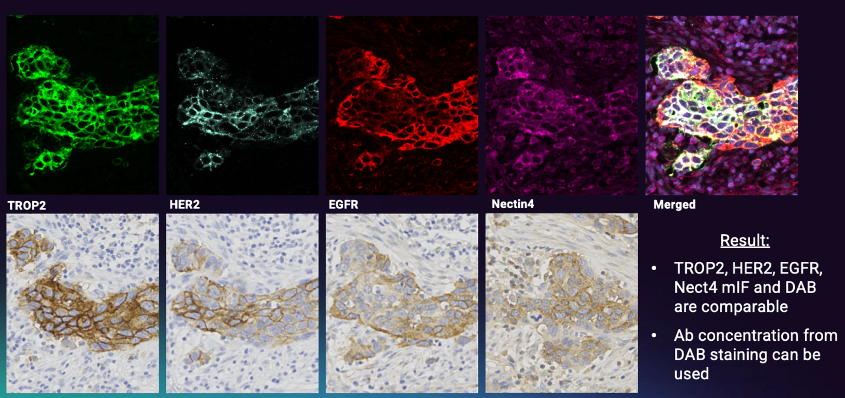
Revolune's reagent platform (RUO) is built around this principle. Rather than requiring proprietary primary antibodies or a specific detection system, the platform is designed to work with the rabbit IgG primary antibodies researchers already trust. The Illuminate-P4-R kit, for example, allows up to four rabbit IgG primary antibodies to be pre-labeled with Revolune Connectors, then applied as a single antibody cocktail in a one-step incubation. Strong concordance between DAB IHC and multiplex IF results (Figure 1) with Revolune-labeled antibodies means validated panels carry over and concentration transfer from established IHC protocols provides a practical starting point for titration. FFPE multiplex IF that works with your existing reagents is the goal.
What Does a Streamlined Path from IHC to Multiplex IF Actually Look Like?
The Revolune workflow (run manually or automated) reduces the complexity of FFPE multiplex IF to a sequence most IHC-experienced researchers will recognize (Figure 2). After standard deparaffinization and heat-induced epitope retrieval, primary antibodies pre-labeled with Revolune Connectors are combined into a single antibody cocktail and applied simultaneously to the tissue section in a one-hour incubation. Two amplification steps follow using the Revolune Amplifier reagents, a DAPI counterstain is applied, and slides are mounted and imaged. The full staining protocol takes approximately six hours after tissue preparation.
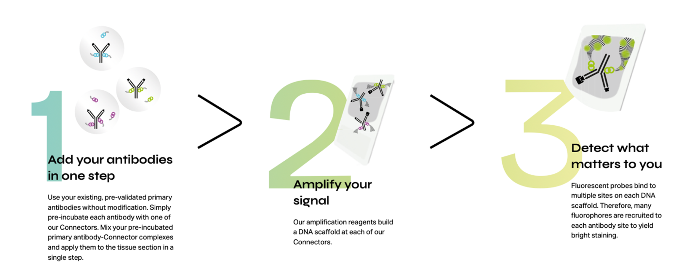
No cyclic stripping. No staining-sequence optimization across multiple rounds. No requirement for specialized imaging hardware beyond a standard fluorescence microscope. For labs that have invested in TSA-based multiplexing workflows, the reduction in hands-on time (6 vs 12 hours) and protocol complexity is meaningful. For labs running single-plex IHC who want to move into spatial proteomics, the transition is easier than ever with the Revolune workflow.
What Does Multiplex IF Enable That Single-Plex IHC Cannot?
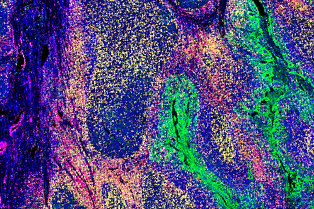
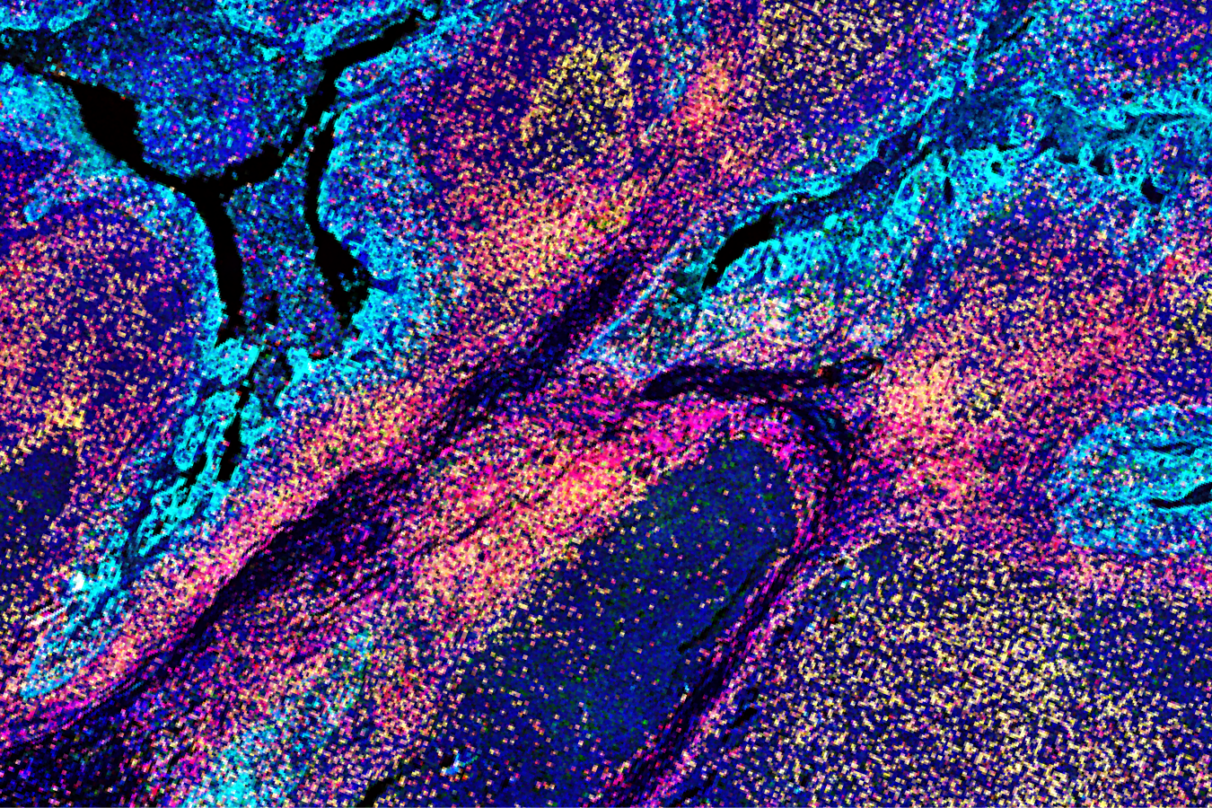
The practical answer is co-expression data at the single-cell level, in intact tissue. Multiplexing same-species primary antibodies in a single staining round means you can simultaneously visualize four biomarkers on one tissue section, preserving spatial relationships between cell populations. For immuno-oncology applications, that means characterizing immune cell phenotypes within the tumor microenvironment in a single experiment rather than across multiple serial sections.
Multiplex IF also supports semi-quantitative to quantitative analysis depending on downstream image analysis approach, enabling comparisons across samples, time points, and treatment arms that single-marker chromogenic data cannot support at the same resolution. As AI-powered tissue analysis tools continue to mature, spatially resolved multiplex IF data becomes the input those tools are designed to use.
Frequently Asked Questions
What is the difference between IHC and multiplex IF?
Standard IHC detects a single protein target per tissue section using a chromogenic reporter. Multiplex IF detects four or more targets simultaneously on one FFPE section using fluorescent reporters, preserving tissue architecture and enabling spatial co-expression analysis at the single-cell level.
Can you multiplex with same-species primary antibodies?
Yes. Revolune Connectors pre-label each primary antibody with a distinct fluorescent channel before the antibody cocktail is applied, eliminating secondary antibody crosstalk. This means same-species rabbit IgG primary antibodies can be used together in a single staining step without signal interference.
Is multiplex immunofluorescence quantitative?
Multiplex IF supports semi-quantitative to quantitative analysis depending on imaging hardware calibration and image analysis workflow. It enables relative comparisons of marker expression across cell populations and samples, and when combined with validated image analysis pipelines, can support more rigorous quantification.
Do you need a special microscope for multiplex IF?
No specialized microscope is required for the Revolune workflow. Standard fluorescence microscopes with appropriate filter sets for the 488, 555, 647, and 750 nm channels are sufficient to image Revolune-stained slides.
What is a TSA alternative for multiplex immunofluorescence?
Tyramide signal amplification (TSA staining) has been a mainstay of cyclic multiplex IF workflows, but requires sequential staining rounds, antibody stripping, and extensive optimization. Revolune offers a TSA alternative that achieves multiplexing through a single staining step with built-in signal amplification. For a deeper comparison, see our next post in this series.
Ready to Explore Multiplex IF?
Revolune's multiplex IF platform gives translational researchers, biomarker teams, and core facilities the freedom to continue to use the primary antibodies they trust in a new way: adding single-cell spatial context to their research. If you are considering the transition from IHC to multiplex immunofluorescence, or are looking for a more accessible spatial biology workflow for FFPE samples, we would like to talk.
Learn more about Revolune’s technology or request a demo.
References
- Sater, S. et al. Society for Immunotherapy of Cancer: Standards for Reporting of Multiplex Immunohistochemistry/Immunofluorescence Assays (STORMI). J Immunother Cancer. 2025 Dec 21;13(12):e012280. doi: 1136/jitc-2025-012280
.webp)