How to Design a Multiplex Immunofluorescent Panel: A Practical mIF Guide

Key takeaways
- Start with the biological question, not the maximum plex count. Every biomarker should contribute to answer your biological question.
- Validate and titrate each antibody in single-plex before multiplexing. Select the lowest antibody concentration that produces the expected staining pattern with minimal background.
- Assign fluorophores according to marker abundance, tissue autofluorescence, and co-localization. Lower-abundance targets generally need more favourable detection channels, while highly expressed targets offer greater flexibility.
- Build the panel iteratively and verify that multiplexing has not changed marker performance. Compare multiplex staining with established single-plex IF or chromogenic IHC and use marker-drop controls to identify interference.
- Match the validation strategy to the intended use. Exploratory, translational, and clinical-facing assays require different levels of testing across runs, reagent lots, operators, instruments, and sites.
Introduction
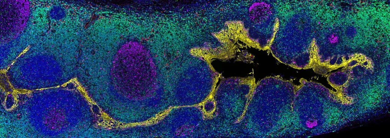
Designing a robust multiplex immunofluorescence, or mIF, panel requires more than selecting a set of biologically interesting biomarkers. Successful mIF panel design combines a clearly defined biological question with validated antibodies, appropriate antibody concentrations, strategic fluorophore assignment, and fit-for-purpose controls.
This practical guide explains how to design and validate a multiplex immunofluorescence panel, from biomarker and antibody selection through single-plex optimization, fluorophore pairing, marker-drop controls, and reproducibility testing.
What Biological Question Should Your mIF Panel Answer?
Before selecting a single antibody, define the scientific question the assay needs to answer.
Ask yourself:
- What decision should the assay support?
- Which cell populations need to be identified?
- Which biomarkers are essential versus exploratory?
A useful rule: If removing a marker would not change your interpretation, you probably do not need that marker.
How Should You Select Antibodies for an mIF Panel?
Begin by identifying antibodies with:
- Proven specificity
- Robust staining patterns
- Published validation data (such as knockout or knockdown validation, orthogonal methods, expected staining pattern in positive and negative control tissues)
- Compatibility with your tissue type
- Compatibility with your staining workflow
Confirm the expected staining pattern using appropriate positive and negative controls. A multi-tissue microarray can be useful for assessing tissue specificity and identifying unexpected staining across tissue types.
Why Should Every Antibody Be Optimized in Single-Plex First?
Multiplex success starts with high-quality single-marker assays. A good practice is to establish each biomarker individually using chromogenic IHC or single-plex IF before integrating it into a multiplex panel.
Each antibody should be titrated independently to identify the optimal signal-to-background ratio. During optimization, evaluate:
- Signal intensity
- Cellular and subcellular localization
- Background staining
- Reproducibility
The goal is not the strongest possible signal. Instead, aim for the lowest concentration that produces a biologically accurate staining pattern with minimal background. Comparison of mIF and DAB staining patterns is a powerful way to verify that multiplexing has not altered biological interpretation.
How Should Biomarkers Be Paired with Fluorophores?
Fluorophore assignment is one of the most critical steps in panel design.
As a general rule:
- High-expression biomarkers can be paired with dimmer fluorophores
- Low-expression biomarkers should be paired with brighter fluorophores
This approach helps balance signal levels across the panel and improves the detectability of lower-abundance targets. Fluorophore assignment should also consider tissue autofluorescence, detector sensitivity, filter compatibility, and potential channel crosstalk.
The recommendations in the table below provide a starting point for biomarker-to-Revolune Connector assignment with Revolune Illuminate reagents. Optimal assignments remain dependent on tissue type, fixation, marker abundance, antibody performance, imaging settings, and the available detection channels.
*Relative ratings are based on internal assay-development experience and are intended as starting guidance, not as universal performance specifications.
How Do Co-Localization and Cellular Localization Affect Panel Design?
Biomarkers that frequently co-localize should ideally not be assigned to spectrally adjacent channels.
For example:
- Two membrane markers expressed on the same cell population may be difficult to discriminate if assigned to neighboring spectral regions
- Markers with clearly distinct subcellular localization may be easier to separate during image analysis
Where possible, avoid assigning strongly co-expressed or spatially overlapping biomarkers to channels with a high risk of signal crosstalk. This can simplify quality control and downstream image analysis.
What Are Marker-Drop Controls and Why Are They Important?
A marker-drop control is a multiplex staining condition in which one primary antibody is omitted while the remaining panel and detection workflow are kept unchanged. Comparing the complete panel with marker-drop conditions can help identify unexpected signal in the omitted channel and assess whether removal of one marker changes the apparent performance of the remaining channels.
Drop testing helps reveal:
- Non-specific signal in an omitted channel
- Signal bleed-through or channel crosstalk
- Detection-reagent interactions
How Much Validation Does an mIF Panel Need?
Validation should be matched to the assay’s intended use. An exploratory research panel may primarily require biological plausibility and within-run controls. A panel intended for larger translational studies may additionally require predefined acceptance criteria and assessments across runs, reagent lots, operators, instruments, and sites. Requirements for clinical use should be established within the applicable quality and regulatory framework.
How Does the Detection Chemistry Affect Panel Design?
The detection chemistry used in a multiplex assay has a direct impact on panel development complexity. Some multiplex workflows require sequential staining, antibody stripping cycles, species matching, or predefined antibody reagents. These requirements can make panel optimization time-consuming, particularly when markers need to be added, removed, or reassigned during development.
With Revolune's Connector-based workflow, panel design follows a modular approach. Each primary antibody is paired with a Revolune Connector independently before multiplex development. Biomarkers can therefore be reassigned to different fluorophores without rebuilding the entire panel, making it straightforward to optimize fluorophore selection as more information becomes available during assay development.
Another advantage is that all antibody-Connector complexes are combined into a single antibody cocktail and applied simultaneously. Because staining occurs in one step, there is no need to determine an optimal antibody staining order, as is often required in sequential multiplex workflows. This simplifies assay development and reduces the number of variables that need to be investigated during optimization.
The modular design is particularly valuable when developing custom panels. If a biomarker needs to be replaced, added, or moved to a different fluorophore channel, only the affected marker typically requires re-evaluation rather than redevelopment of the entire assay. This enables a more iterative and flexible approach to panel design and can substantially accelerate assay development.
In practice, this means researchers can focus more on the biological question and less on managing technical constraints arising from the multiplex chemistry itself. When choosing a multiplex platform, it is therefore important to consider not only signal quality and plex size, but also how easily panels can be modified, expanded, and transferred between projects.